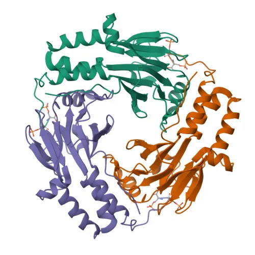
Phosphorylated Antibodies
Phospho-Smad2-S465/S467 Rabbit pAb for WB, ELISA - Q15796
Item Number : CM0000494
Price varies based on specs and customizations
- Application
- WB, ELISA
- Cross Reactivity
- Human, Mouse
- Protein Weight
- 52 kDa

Shipping:
Contact us to get shipping details Enjoy On-time Dispatch Guarantee.
Why Choose Us
Easy ordering process, quality products, and dedicated support for your business success.
Core Product Specifications and Parameters
| Parameter | Value |
|---|---|
| Product Name | Phospho-Smad2-S465/S467 Rabbit pAb |
| Remarks/Alias | JV18; LDS6; CHTD8; MADH2; MADR2; JV18-1; hMAD-2; hSMAD2; Phospho-Smad2-S465/S467 |
| Species | Human |
| Gene ID (Human) | 4087 |
| Immunogen | Synthetic Peptide|A synthetic phosphorylated peptide around S465 & S467 of human Smad2 (NP_005892.1) |
| Source | Rabbit |
| Category | Phosphorylated Antibodies |
| Application | WB, ELISA |
| Cross Reactivity | Human, Mouse |
| SWISS | Q15796 |
| Protein Weight | 52 kDa |
| Shipping | Ice bag |
Biological Background: Smad2 Function and Localization
- Smad2, also known as MADH2, MADR2, or JV18-1, is a member of the receptor-regulated SMAD (R-SMAD) family, acting as an intracellular signal transducer downstream of TGF-beta and activin receptors.
- Upon phosphorylation at S465 and S467 by type 1 receptor kinases, Smad2 forms a complex with Smad4, translocates to the nucleus, and binds to the TRE element to regulate transcription of TGF-beta-responsive genes.
- Smad2 promotes TGFB1-mediated transcription of odontoblastic differentiation genes in dental papilla cells and positively regulates PDPK1 kinase activity by dissociating it from YWHAQ (14-3-3 protein).
- It may function as a tumor suppressor in colorectal carcinoma (PubMed:8752209). Related references: PMID:8752209
- Subcellular localization is primarily cytoplasmic under basal conditions, but it shuttles to the nucleus upon activation, consistent with its role in signal transduction.
- Post-translational modifications include phosphorylation (critical for activation), acetylation, and ubiquitination (Ubl conjugation). The protein contains a zinc-binding domain and is alternatively spliced.
- Smad2 is highly expressed in skeletal muscle, endothelial cells, heart, and placenta, reflecting its broad role in development and tissue homeostasis.
Experimental Guidance and Technical Tips
- The immunogen is a synthetic phosphorylated peptide encompassing S465 and S467 of human Smad2; thus, the antibody likely recognizes the doubly phosphorylated (active) form. To preserve epitope phosphorylation, add phosphatase inhibitors during sample preparation.
- For Western blot (WB), use a reducing SDS-PAGE and transfer to PVDF; consider optimizing blocking and antibody dilution using relevant cell lysates treated with TGF-beta1 or a phosphatase inhibitor control.
- For ELISA, use a phospho-specific detection system and validate with peptide competition assays to confirm specificity.
- Validate cross-reactivity with mouse samples, as reactivity is predicted; perform a species-specific alignment of the epitope if needed for other species.
CamelBio: Your One-Stop Sourcing Bridge
CamelBio is a comprehensive IVD raw-material sourcing partner, bridging diagnostic manufacturers and research laboratories with high-quality antibodies and ancillary reagents. This phospho-Smad2 antibody supports research in TGF-beta signaling, cancer biology, and developmental signaling pathways. Explore our validated monoclonal and polyclonal antibodies, bulk reagent supply, and custom sourcing for rare targets to accelerate your assay development.
REQUEST A QUOTE
Our professional team will reply to you within one business day. Please feel free to contact us!