Monoclonal Antibodies
PE/Cyanine7 Rabbit anti-Human/Monkey PD-1/CD279 Monoclonal Antibody for FC - Q15116
Item Number : CM0012785
Price varies based on specs and customizations
- Application
- FC
- Cross Reactivity
- Human, Cynomolgus
- Protein Weight
- 32kDa

Shipping:
Contact us to get shipping details Enjoy On-time Dispatch Guarantee.
Why Choose Us
Easy ordering process, quality products, and dedicated support for your business success.
Core Product Specifications and Parameters
| Parameter | Value |
|---|---|
| Product Name | PE/Cyanine7 Rabbit anti-Human/Monkey PD-1/CD279 mAb |
| Remarks/Alias | PD1; PD-1; CD279; SLEB2; hPD-1; hPD-l; hSLE1 |
| Species | Human |
| GeneID (Human) | 5133 |
| GeneID | 5133 |
| Immunogen | Recombinant fusion protein containing a sequence corresponding to amino acids 25-168 of human PD-1/CD279 (NP_005009.2) |
| Source | Rabbit |
| Category | Monoclonal Antibodies |
| Application | FC |
| Cross Reactivity | Human, Cynomolgus |
| SWISS | Q15116 |
| Protein Weight | 32kDa |
| Shipping | Ice bag |
Biological Background: PD-1 Function and Localization
- PD-1 (Programmed cell death protein 1), also known as CD279, is an inhibitory receptor encoded by the PDCD1 gene that plays a critical role in induction and maintenance of immune tolerance. Related references: PMID:21276005, PMID:31754127, PMID:32184441
- It delivers inhibitory signals upon binding to its ligands CD274/PDCD1L1 and CD273/PDCD1LG2, downregulating T-cell responses. Related references: PMID:21276005, PMID:26602187
- Following T-cell receptor (TCR) engagement, PD-1 associates with TCR-CD3 in the immunological synapse and directly inhibits T-cell activation by recruiting PTPN11/SHP-2. Related references: PMID:32184441
- The SHP-2 phosphatase dephosphorylates key TCR proximal signaling molecules, including ZAP70, PRKCQ/PKCθ, and CD247/CD3ζ, thereby suppressing T-cell activation. Related references: PMID:32184441
- The PD-1-mediated inhibitory pathway is exploited by tumors to attenuate anti-tumor immunity; blockade of this pathway reverses exhausted T-cell phenotypes and restores anti-tumor responses, forming the basis of cancer immunotherapy. Related references: PMID:28951311, PMID:22658127, PMID:25034862, PMID:25399552
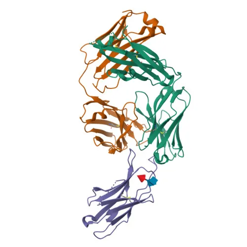
- PD-1 is a single-pass transmembrane glycoprotein localized to the cell membrane and contains an immunoglobulin-like domain, disulfide bonds, and intracellular signaling motifs subject to phosphorylation and ubiquitination.
Experimental Guidance and Technical Tips
- The immunogen used spans the extracellular domain (amino acids 25-168) of human PD-1, suggesting the antibody targets the native cell-surface conformation.
- When using this PE/Cyanine7-conjugated antibody for flow cytometry, ensure proper compensation settings and include appropriate isotype controls to account for non-specific binding.
- Cross-reactivity with cynomolgus monkey (Macaca fascicularis) PD-1 has been observed, potentially enabling translational studies in non-human primate models.
- It is advisable to titrate the antibody and validate its performance in the relevant sample system, as optimal conditions may vary depending on cell type and experimental setup.
CamelBio: Your One-Stop Sourcing Bridge
CamelBio offers diagnostic manufacturers, labs, and research institutes a streamlined one-stop channel for IVD raw materials, covering validated antibody pairs, optimized monoclonal and polyclonal antibodies, bulk ancillary reagents, and rare target sourcing. For PD-1/immune checkpoint research and immunotherapy development, CamelBio can supply this conjugated anti-PD-1 antibody alongside complementary reagents to support your discovery and translational workflows. Our integrated supply model helps you move from concept to clinic with reliable material access and expert consultation.
REQUEST A QUOTE
Our professional team will reply to you within one business day. Please feel free to contact us!