Monoclonal Antibodies
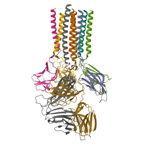
PE Mouse anti-Human TCR Vδ2 Monoclonal Antibody for FC - P04234
Item Number : CM0011013
Price varies based on specs and customizations
- Application
- FC
- Cross Reactivity
- Human
- Protein Weight
- 19kDa

Shipping:
Contact us to get shipping details Enjoy On-time Dispatch Guarantee.
Why Choose Us
Easy ordering process, quality products, and dedicated support for your business success.
Core Product Specifications and Parameters
| Parameter | Value |
|---|---|
| Product Name | PE Mouse anti-Human TCR Vδ2 mAb |
| Remarks/Alias | TCRD; TRD@; TCRDV1 |
| Species | Human |
| GeneID | 6964 |
| Immunogen | Recombinant protein of human TCR Vδ2 |
| Source | Mouse |
| Category | Monoclonal Antibodies |
| Application | FC |
| Cross Reactivity | Human |
| SWISS | P04234 |
| Protein Weight | 19kDa |
| Shipping | Ice bag |
Biological Background: CD3 Delta Chain Function and Localization
- The CD3D gene encodes the CD3 delta chain, a critical component of the T-cell receptor-CD3 complex on T-lymphocytes, playing an essential role in adaptive immune responses.
- CD3 delta, together with CD3E, CD3G, and CD247, transmits activation signals across the plasma membrane after TCR engagement by peptide-MHC complexes on antigen-presenting cells.
- The cytoplasmic domain contains immunoreceptor tyrosine-based activation motifs (ITAMs) that are phosphorylated by Src kinases LCK and FYN, initiating downstream signaling cascades.
Related references: PMID:2470098 - CD3D is indispensable for thymocyte differentiation; it ensures proper TCR-CD3 complex assembly and surface expression, and physically links the TCR to CD4 or CD8 co-receptors, a process required for positive selection of T-cells.
Related references: PMID:12215456 - Subcellularly, the CD3 delta chain localizes predominantly to the cell membrane as a single-pass type I membrane protein.
- It is primarily expressed on T-lymphocytes as part of the mature TCR-CD3 complex and is also detected in fetal natural killer (NK) cells.
- Post-translational modifications include glycosylation and phosphorylation; the protein features a signal peptide, an extracellular immunoglobulin-like domain, a transmembrane helix, and a cytoplasmic ITAM-containing region.
Experimental Guidance and Technical Tips
- Since the immunogen is recombinant TCR Vδ2, verify that the antibody recognizes the native conformation on live cell surfaces during flow cytometry staining.
- Titrate the antibody to determine the optimal working concentration for your specific cell type and experimental conditions.
- Include proper isotype controls and, where possible, fluorescence-minus-one (FMO) controls to set accurate gating boundaries in multicolor panels.
- As a PE-conjugated reagent, handle and store the antibody protected from light to prevent photobleaching and signal loss.
- The reported cross reactivity is human; if testing non-human primate or other species, perform an independent validation.
- For consistent results, standardize fixation and permeabilization steps if intracellular staining is attempted, although the antibody is intended for surface staining.
CamelBio: Your One-Stop Sourcing Bridge
CamelBio offers a comprehensive portfolio of IVD raw materials and technical services for diagnostic and research applications, including validated antibody pairs, optimized monoclonal/polyclonal antibodies, and ancillary reagents. If your project involves T-cell immunophenotyping or gamma/delta T cell biology, we supply a range of fluorescently labeled antibodies against TCR chains and CD3 components to meet your assay requirements. Contact us to streamline your sourcing and accelerate development from concept to clinic.
REQUEST A QUOTE
Our professional team will reply to you within one business day. Please feel free to contact us!