Polyclonal Antibodies
HLA-DPB1 Rabbit pAb for WB, IHC-P, ELISA - P04440
Item Number : CM0013130
Price varies based on specs and customizations
- Application
- WB, IHC-P, ELISA
- Cross Reactivity
- Human, Mouse, Rat
- Protein Weight
- 29 kDa

Shipping:
Contact us to get shipping details Enjoy On-time Dispatch Guarantee.
Why Choose Us
Easy ordering process, quality products, and dedicated support for your business success.
Core Product Specifications and Parameters
| Specification | Details |
|---|---|
| Product Name | HLA-DPB1 Rabbit pAb |
| Remarks/Alias | DPB1; HLA-DP; HLA-DPB; HLA-DP1B; HLA-DPB1 |
| Species | Human |
| Gene ID | 3115 |
| Immunogen | Recombinant fusion protein containing a sequence corresponding to amino acids 30-225 of human HLA-DPB1 (NP_002112.3) |
| Source | Rabbit |
| Category | Polyclonal Antibodies |
| Application | WB, IHC-P, ELISA |
| Cross Reactivity | Human, Mouse, Rat |
| SWISS | P04440 |
| Protein Weight | 29 kDa |
| Shipping | Ice bag |
Biological Background: HLA-DPB1 Function and Localization
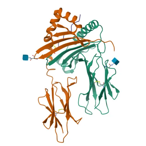
- HLA-DPB1 encodes the beta chain of the MHC class II molecule HLA-DP, a cell surface receptor essential for adaptive immune responses.
- It presents peptides derived from exogenous antigens that have been internalized via endocytosis or autophagy to CD4+ T helper cells, triggering immune activation.
- The peptide-binding cleft accommodates peptides of 10–30 residues, generated mainly by lysosomal proteolysis in antigen-presenting cells (APCs).
- Expressed primarily on professional APCs such as B cells, dendritic cells, and macrophages, as well as on gastrointestinal epithelial cells.
- Intracellularly, HLA-DPB1 traverses the endoplasmic reticulum, Golgi, and endosomal/lysosomal compartments before reaching the cell membrane.
- The protein undergoes glycosylation and contains a disulfide bond, and its stable surface expression depends on interaction with accessory molecules CD74 and HLA-DM during peptide loading.
Experimental Guidance and Technical Tips
- The immunogen corresponds to the extracellular domain (aa 30-225) of human HLA-DPB1, suggesting potential reactivity against both native and denatured protein forms.
- For Western blot, consider using lysates from human B-cell lines or spleen tissue to detect the ~29 kDa band; validate in your system with appropriate controls.
- For IHC-P, human lymphoid organs (tonsil, lymph node) are suitable positive controls; optimize antigen retrieval and blocking conditions.
- Evaluate cross-reactivity in mouse and rat samples, as sequence homology may affect recognition; confirm with parallel human controls.
- ELISA performance may depend on coating conditions and antibody pair compatibility; empirical testing is recommended.
CamelBio: Your One-Stop Sourcing Bridge
CamelBio streamlines IVD raw-material procurement for diagnostic manufacturers and research labs, offering this HLA-DPB1 polyclonal antibody alongside a broad inventory of immunology-related reagents. Whether you need validated antibody pairs, optimized mono- or polyclonal antibodies, bulk ancillary reagents, or customized sourcing for rare targets like MHC class II proteins, CamelBio serves as your integrated supply partner from discovery to clinical application.
REQUEST A QUOTE
Our professional team will reply to you within one business day. Please feel free to contact us!