Monoclonal Antibodies
Anti-VDR Rabbit Monoclonal Antibody for WB, IP, IF/ICC, IF-P, IHC-P, ELISA - P11473
Item Number : CM0004245
Price varies based on specs and customizations
- Application
- WB, IP, IF/ICC, IF-P, IHC-P, ELISA
- Cross Reactivity
- Human, Mouse, Rat
- Protein Weight
- 48kDa

Shipping:
Contact us to get shipping details Enjoy On-time Dispatch Guarantee.
Why Choose Us
Easy ordering process, quality products, and dedicated support for your business success.
Core Product Specifications and Parameters
| Parameter | Value |
|---|---|
| Product Name | VDR Rabbit mAb |
| Remarks/Alias | NR1I1; PPP1R163; VDR |
| Species | Human |
| GeneID | 7421 |
| Immunogen | Synthetic peptide |
| Source | Rabbit |
| Category | Monoclonal Antibodies |
| Application | WB, IP, IF/ICC, IF-P, IHC-P, ELISA |
| Cross Reactivity | Human, Mouse, Rat |
| SWISS | P11473 |
| Protein Weight | 48kDa |
| Shipping | Ice bag |
Biological Background: VDR Function and Localization
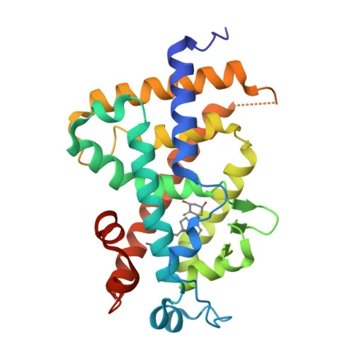
- VDR (vitamin D3 receptor) is a nuclear receptor for calcitriol, the active form of vitamin D3, mediating its genomic actions on target cells. Related references: PMID:10678179, PMID:15728261, PMID:16913708
- Upon ligand binding, VDR heterodimerizes with retinoid X receptor (RXR) and binds vitamin D response elements (VDREs) in DNA to regulate transcription of target genes. Related references: PMID:28698609
- Plays a central role in calcium and phosphate homeostasis, bone metabolism, and cell differentiation.
- Also functions as a receptor for the secondary bile acid lithocholic acid (LCA) and its metabolites, linking vitamin D signaling to bile acid detoxification. Related references: PMID:12016314, PMID:32354638
- Subcellular localization primarily in the nucleus, but also detected in the cytoplasm; ligand binding promotes nuclear translocation.
- Contains two zinc-finger motifs essential for DNA binding, and undergoes post-translational modifications such as ubiquitination and phosphorylation.
- Alternative splicing produces multiple isoforms; genetic variants in VDR are associated with disorders like vitamin D-resistant rickets.
Experimental Guidance and Technical Tips
- For Western blot, expect a major band around 48 kDa; validate in relevant cell or tissue lysates expressing VDR.
- In immunoprecipitation and ELISA, optimize antibody concentration and buffer conditions for the target protein from human, mouse, or rat samples.
- For immunofluorescence and immunohistochemistry, confirm specificity using positive control tissues (e.g., kidney, intestine) and ensure proper fixation/permeabilization protocols.
- The immunogen (N-terminal 1–100 amino acids) may recognize multiple VDR isoforms; consider this when interpreting results across different applications.
CamelBio: Your One-Stop Sourcing Bridge
CamelBio serves as a comprehensive procurement partner for diagnostic manufacturers and research institutes, offering a verified portfolio of IVD raw materials including validated monoclonal and polyclonal antibodies, antibody pairs, and bulk ancillary reagents. For vitamin D receptor-related research and assay development, CamelBio streamlines access to high-quality anti-VDR antibodies and rare target raw materials, supporting projects from early discovery through clinical application. Our supply capabilities help you reliably source optimized reagents tailored to your workflow requirements.
REQUEST A QUOTE
Our professional team will reply to you within one business day. Please feel free to contact us!