Polyclonal Antibodies
Anti-UBA1 Polyclonal Antibody for WB, IHC-P, IF/ICC, ELISA - P22314
Item Number : CM0017867
Price varies based on specs and customizations
- Application
- WB, IHC-P, IF/ICC, ELISA
- Cross Reactivity
- Human, Mouse, Rat
- Protein Weight
- 118kDa

Shipping:
Contact us to get shipping details Enjoy On-time Dispatch Guarantee.
Why Choose Us
Easy ordering process, quality products, and dedicated support for your business success.
Core Product Specifications and Parameters
| Parameter | Value |
|---|---|
| Product Name | UBA1 Rabbit pAb |
| Remarks/Alias | A1S9; A1ST; GXP1; UBE1; A1S9T; AMCX1; POC20; SMAX2; UBA1A; UBE1X; VEXAS; CFAP124; UBA1 |
| Species | Human |
| Gene ID | 7317 |
| Immunogen | Recombinant fusion protein containing a sequence corresponding to amino acids 300-600 of human UBA1 (NP_695012.1) |
| Source | Rabbit |
| Category | Polyclonal Antibodies |
| Application | WB, IHC-P, IF/ICC, ELISA |
| Cross Reactivity | Human, Mouse, Rat |
| SWISS | P22314 |
| Protein Weight | 118kDa |
| Shipping | Ice bag |
Biological Background: UBA1 Function and Localization
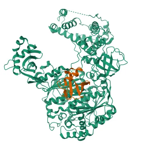
- UBA1 (Ubiquitin-like modifier-activating enzyme 1), also known as Ubiquitin-activating enzyme E1 or Protein A1S9, is the primary E1 enzyme responsible for initiating ubiquitin conjugation, a critical step in marking proteins for degradation via the ubiquitin-proteasome system. Related references: PMID:1447181 PMID:1606621 PMID:33108101
- Specifically, UBA1 activates ubiquitin by adenylating its C-terminal glycine with ATP, then forms a thioester bond between ubiquitin and its own catalytic cysteine, enabling transfer to E2 enzymes. Related references: PMID:1447181
- UBA1 plays an essential role in DNA damage response by promoting the formation of radiation-induced foci and the recruitment of repair proteins like TP53BP1 and BRCA1 to damage sites, thereby facilitating timely DNA repair and replication stress response. Related references: PMID:22456334
- The protein localizes to the cytoplasm, nucleus, and mitochondrion, reflecting its broad cellular function in protein homeostasis and signaling.
- UBA1 is ubiquitously expressed, with detection in erythrocytes at the protein level, and is evolutionarily conserved across human, mouse, and rat.
- Post-translational modifications include acetylation and phosphorylation, which may regulate its activity; the gene is also associated with disease variants linked to neurodegeneration.
- Keywords from UniProt highlight its roles as an ATP-binding ligase with repeated domains, involvement in Ubl conjugation pathways, and its identification in proteomic studies.
Experimental Guidance and Technical Tips
- The immunogen corresponds to amino acids 300-600 of human UBA1, a region that is conserved across human, mouse, and rat, supporting the observed cross-reactivity. For species-specific detection, validate in your model system.
- For Western blotting (WB), the predicted molecular weight is ~118 kDa; ensure adequate gel resolution and transfer conditions for high-molecular-weight proteins.
- For immunohistochemistry (IHC-P) and immunofluorescence (IF/ICC), consider antigen retrieval and appropriate fixation/permeabilization protocols tailored to your sample type.
- For ELISA, use as capture or detection antibody after confirming binding affinity to native versus recombinant UBA1.
- As a polyclonal antibody, it may detect multiple isoforms or degradation products; verify specificity with appropriate controls (e.g., knockdown or knockout lysates).
CamelBio: Your One-Stop Sourcing Bridge
CamelBio is your one-stop sourcing partner for IVD raw materials, supporting diagnostic manufacturers, research labs, and institutes from concept to clinic. With this anti-UBA1 polyclonal antibody, we empower your research into ubiquitin-proteasome pathways and related diseases. Beyond individual reagents, CamelBio offers validated antibody pairs, bulk ancillary reagents, and custom sourcing for rare targets—streamlining your supply chain and accelerating assay development.
REQUEST A QUOTE
Our professional team will reply to you within one business day. Please feel free to contact us!