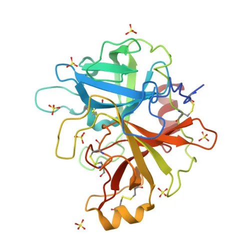
Polyclonal Antibodies
Anti-ST14 Polyclonal Antibody for WB, ELISA - Q9Y5Y6
Item Number : CM0024812
Price varies based on specs and customizations
- Application
- WB, ELISA
- Cross Reactivity
- Human, Mouse
- Protein Weight
- 95kDa

Shipping:
Contact us to get shipping details Enjoy On-time Dispatch Guarantee.
Why Choose Us
Easy ordering process, quality products, and dedicated support for your business success.
Core Product Specifications and Parameters
| Parameter | Value |
|---|---|
| Product Name | ST14 Rabbit pAb |
| Remarks/Alias | HAI, CAP3, MTSP1, SNC19, ARCI11, MT-SP1, PRSS14, TADG15, TMPRSS14, ST14 |
| Species | Human |
| GeneID (Human) | 6768 |
| GeneID | 6768 |
| Immunogen | Recombinant fusion protein containing a sequence corresponding to amino acids 566-855 of human ST14 (NP_068813.1) |
| Source | Rabbit |
| Category | Polyclonal Antibodies |
| Application | WB, ELISA |
| Cross Reactivity | Human, Mouse |
| SWISS | Q9Y5Y6 |
| Protein Weight | 95kDa |
| Shipping | Ice bag |
Biological Background: ST14 Function and Localization
- ST14 encodes suppressor of tumorigenicity 14 protein, also known as matriptase, a trypsin-like serine protease that cleaves substrates at Arg or Lys residues. Related references: PMID:10373424
- It plays a key role in terminal keratinocyte differentiation by activating prostasin (PRSS8) and processing filaggrin (FLG). Related references: PMID:18843291
- ST14 proteolytically cleaves and activates TMPRSS13, a transmembrane serine protease. Related references: PMID:28710277
- The protein is a membrane-anchored serine protease with a signal-anchor transmembrane domain, localizing to the plasma membrane.
- ST14 contains multiple disulfide bonds and glycosylation sites, and undergoes proteolytic processing for activation.
- Mutations in ST14 are associated with autosomal recessive ichthyosis and hypotrichosis, highlighting its biological significance in epithelial tissues.
Experimental Guidance and Technical Tips
- The immunogen is a recombinant fusion protein covering the C-terminal region (amino acids 566-855), which may recognize both full-length ST14 and its C-terminal cleavage products.
- For Western blot, consider using whole-cell lysates from human keratinocyte cell lines (e.g., HaCaT) or tumor cell lines known to express ST14; the predicted molecular weight is 95 kDa, but additional bands may appear due to proteolytic activation.
- For ELISA, validate the antibody with recombinant ST14 protein or a peptide spanning the immunogen region, and titrate the antibody to determine optimal working concentration.
- As a polyclonal antibody, lot-to-lot consistency should be verified for critical assays; consider testing cross-reactivity with mouse samples as indicated.
CamelBio: Your One-Stop Sourcing Bridge
CamelBio serves as a comprehensive one-stop sourcing partner for IVD raw materials, connecting diagnostic manufacturers and research labs with validated antibodies, bulk reagents, and rare target materials. Our anti-ST14 polyclonal antibody supports research into protease function and epithelial biology, and we can further assist with matched antibody pairs, optimized monoclonals, and customized sourcing to accelerate your diagnostic development.
REQUEST A QUOTE
Our professional team will reply to you within one business day. Please feel free to contact us!