Monoclonal Antibodies
Anti-SARS-CoV Spike RBD Monoclonal Antibody for WB, ELISA - P59594
Item Number : CM0006970
Price varies based on specs and customizations
- Application
- WB, ELISA
- Cross Reactivity
- SARS-CoV
- Protein Weight
- 139kDa

Shipping:
Contact us to get shipping details Enjoy On-time Dispatch Guarantee.
Why Choose Us
Easy ordering process, quality products, and dedicated support for your business success.
Core Product Specifications and Parameters
| Parameter | Value |
|---|---|
| Product Name | SARS-CoV Spike RBD Rabbit mAb |
| Remarks/Alias | E2; SARS-CoV Spike RBD |
| Gene ID | 1489668 |
| Immunogen | Synthetic peptide corresponding to a sequence within amino acids 350-450 of coronavirus SARS-CoV Spike RBD (NP_828851.1) |
| Source | Rabbit |
| Category | Monoclonal Antibodies |
| Application | WB, ELISA |
| Cross Reactivity | SARS-CoV |
| SWISS | P59594 |
| Protein Weight | 139kDa |
| Shipping | Ice bag |
Biological Background: SARS-CoV Spike RBD Function and Localization
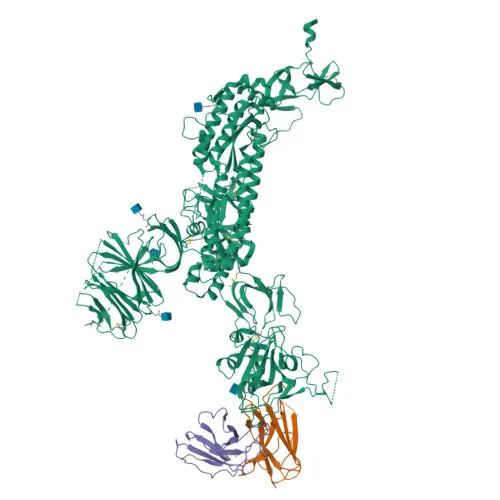
- The Spike glycoprotein, also known as E2 or peplomer protein, is a 139 kDa class I viral fusion protein encoded by the S gene of SARS-CoV (UniProt P59594).
- It mediates viral attachment to host cells by binding to receptors such as human ACE2 and CLEC4M/DC-SIGNR, triggering internalization into endosomes (By similarity).
- Following endocytosis, cathepsin CTSL proteolysis unmasks the fusion peptide in the S2 subunit, activating membrane fusion between the viral envelope and host endosomal membrane.
- The protein also down-regulates host tetherin (BST2) via lysosomal degradation, counteracting innate antiviral activity.
- Subcellular localization includes the virion membrane, host endoplasmic reticulum-Golgi intermediate compartment membrane, and host cell membrane.
- Structural features include a signal peptide, coiled coil heptad repeats, disulfide bonds, a transmembrane helix, and palmitoylation, all essential for its fusogenic function.
- Keywords: Viral envelope protein, virus entry, host-virus interaction, immunoevasion, fusion of virus membrane with host membrane.
Experimental Guidance and Technical Tips
- The immunogen corresponds to residues 350–450 of the Spike RBD; the resulting antibody is expected to recognize the native RBD in Western blot under reducing or non-reducing conditions.
- For ELISA, consider this monoclonal antibody as a capture or detection reagent; matrix effects and dynamic range should be empirically determined.
- Validate specificity and cross-reactivity in the relevant experimental system (e.g., SARS-CoV infected cell lysates or recombinant RBD proteins).
- No cross-reactivity data are provided for other coronaviruses; testing is recommended if broad reactivity is required.
CamelBio: Your One-Stop Sourcing Bridge
CamelBio streamlines IVD raw-material procurement for SARS-CoV spike research and diagnostic development. We offer validated monoclonal antibodies like this anti-RBD clone, alongside optimized antibody pairs, bulk ancillary reagents, and sourcing support for rare targets. Reach out to explore how CamelBio can accelerate your project from concept to clinic.
REQUEST A QUOTE
Our professional team will reply to you within one business day. Please feel free to contact us!