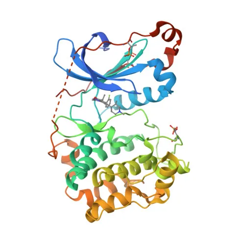
Phosphorylated Antibodies
Anti-Phospho-PKC alpha-T638 Rabbit mAb for WB, ELISA - P17252
Item Number : CM0001410
Price varies based on specs and customizations
- Application
- WB, ELISA
- Cross Reactivity
- Human
- Protein Weight
- 77kDa

Shipping:
Contact us to get shipping details Enjoy On-time Dispatch Guarantee.
Why Choose Us
Easy ordering process, quality products, and dedicated support for your business success.
Core Product Specifications and Parameters
| Parameter | Value |
|---|---|
| Product Name | Phospho-PKC alpha-T638 Rabbit mAb |
| Remarks/Alias | AAG6; PKCA; PRKACA; PKCI+/-; PKCalpha; PKC-alpha; Phospho-PKC alpha-T638 |
| Species | Human |
| Gene ID (Human) | 5578 |
| Gene ID | 5578 |
| Immunogen | Synthetic peptide|A synthetic phosphorylated peptide around T638 of human PKC alpha. |
| Source | Rabbit |
| Category | Phosphorylated Antibodies |
| Application | WB, ELISA |
| Cross Reactivity | Human |
| SWISS | P17252 |
| Protein Weight | 77kDa |
| Shipping | Ice bag |
Biological Background: PKC alpha Function and Localization
- Protein kinase C alpha type (PKCα) is a calcium-activated, phospholipid- and diacylglycerol (DAG)-dependent serine/threonine-protein kinase that regulates diverse cellular processes including proliferation, apoptosis, differentiation, migration, adhesion, and angiogenesis.
- PKCα operates in both positive and negative regulation of cell cycle progression; it can promote growth via RAF1/MAPK/ERK signaling or induce cell cycle arrest through RB1 hyperphosphorylation and CDK inhibitor induction (CDKN1A, CDKN1B).
- The kinase exhibits anti-apoptotic functions in cancer cells by suppressing p53/TP53-mediated IGFBP3 activation and phosphorylating BCL2, thereby contributing to tumor promotion in gliomas and breast cancers.
- PKCα plays a critical role in cardiovascular biology: it negatively regulates myocardial contractility, drives cardiomyocyte hypertrophy via ERK1/2 signaling, and promotes angiogenesis by enhancing endothelial cell migration and VEGFA-induced proliferation.
- In platelet function, PKCα mediates CD36/GP4-dependent granule release and integrin ITGA2B-ITGB3 activation through the RAP1GAP pathway, contributing to thrombus formation.
- The kinase is involved in immune responses, including macrophage differentiation and chemokine-induced T cell migration; it phosphorylates DOCK8 to regulate CDC42 activation. Related references: PMID:28028151
- Subcellular localization of PKCα is dynamic: it is found in the cytoplasm, cell membrane, mitochondrial membrane, and nucleus, depending on activation status and stimuli.
- PKCα phosphorylates a wide array of substrates, including TNNT2/CTNT (regulating cardiac contractility), KLHL3 (modulating WNK4 stability in response to angiotensin II), and LRRK1 (involved in intracellular trafficking). Related references: PMID:25313067 PMID:36040231
Experimental Guidance and Technical Tips
- The immunogen is a synthetic phosphopeptide spanning Thr638 of human PKCα; antibody specificity for the phosphorylated form should be validated by comparing signals in phosphatase-treated versus untreated samples.
- For Western blot, consider using low percentage SDS-PAGE gels to resolve the 77 kDa protein, and include appropriate loading controls alongside non‑phospho‑specific PKCα antibodies to assess total protein levels.
- In ELISA applications, investigate coating antigen concentrations and blocking conditions empirically; a phosphopeptide competition assay may help confirm phospho‑specific binding.
- Because PKCα activation is transient and stimulus‑dependent, optimize cell stimulation protocols (e.g., PMA, growth factors) and ensure timely sample preparation with phosphatase inhibitors.
CamelBio: Your One-Stop Sourcing Bridge
CamelBio is a dedicated one-stop IVD raw‑material sourcing partner, supporting diagnostic manufacturers, laboratories, and research institutes with validated antibody pairs, bulk ancillary reagents, and rare‑target custom development. This anti‑phospho‑PKCα‑T638 antibody addresses a key node in oncology, cardiovascular, and inflammation signal transduction research. We streamline procurement from concept to clinic, offering reliable monoclonal antibodies and tailored raw‑material solutions for high‑sensitivity assay development.
REQUEST A QUOTE
Our professional team will reply to you within one business day. Please feel free to contact us!