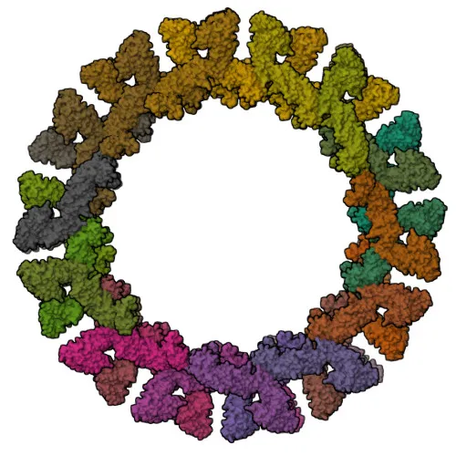
Monoclonal Antibodies
Anti-OPA1 Rabbit Monoclonal Antibody for WB, IP, IF/ICC, IF-P, ELISA - O60313
Item Number : CM0009704
Price varies based on specs and customizations
- Application
- WB, IP, IF/ICC, IF-P, ELISA
- Cross Reactivity
- Human, Mouse, Rat
- Protein Weight
- 108-118 kDa

Shipping:
Contact us to get shipping details Enjoy On-time Dispatch Guarantee.
Why Choose Us
Easy ordering process, quality products, and dedicated support for your business success.
Core Product Specifications and Parameters
| Parameter | Value |
|---|---|
| Product Name | OPA1 Rabbit mAb |
| Remarks/Alias | NPG; NTG; MGM1; BERHS; largeG; MTDPS14 |
| Species | Human |
| GeneID (Human) | 4976 |
| GeneID | 4976 |
| Immunogen | Recombinant fusion protein containing a sequence corresponding to amino acids 210-573 of human OPA1 (NP_056375.2) |
| Source | Rabbit |
| Category | Monoclonal Antibodies |
| Application | WB, IP, IF/ICC, IF-P, ELISA |
| Cross Reactivity | Human, Mouse, Rat |
| SWISS | O60313 |
| Protein Weight | 108-118 kDa |
| Shipping | Ice bag |
Biological Background: OPA1 Function and Localization
- OPA1 is a dynamin-related GTPase essential for normal mitochondrial morphology. It exists as two major forms: a long transmembrane form (L-OPA1) anchored to the inner mitochondrial membrane and a short soluble form (S-OPA1) generated by proteolytic cleavage in the intermembrane space.
Related references: PMID:31922487, PMID:32228866, PMID:33237841 - L-OPA1 and S-OPA1 cooperate to mediate mitochondrial inner membrane fusion, a process critical for mitochondrial network maintenance and respiratory chain function.
Related references: PMID:16778770, PMID:17709429, PMID:20185555, PMID:31922487, PMID:37612504, PMID:37612506 - The equilibrium between L-OPA1 and S-OPA1 is tightly regulated: excessive S-OPA1 production by OMA1 following mitochondrial depolarization disrupts fusion and alters cristae morphology.
Related references: PMID:20038677, PMID:31922487 - OPA1 plays a role in mitochondrial genome maintenance; isoforms containing the alternative exon 4b are required for anchoring mitochondrial nucleoids to the inner membrane.
Related references: PMID:18158317, PMID:20974897 - OPA1 is involved in cristae remodeling and facilitates cytochrome c release during apoptosis, linking mitochondrial dynamics to programmed cell death pathways.
- Highly expressed in retina, with additional expression in brain, testis, heart, and skeletal muscle; mutations in OPA1 are associated with optic atrophy, deafness, and neurodegenerative disorders.
- OPA1 localizes to the mitochondrial inner membrane and intermembrane space.
Experimental Guidance and Technical Tips
- The immunogen used for this antibody is a recombinant fusion protein corresponding to amino acids 210-573 of human OPA1 (NP_056375.2). When performing Western blot, anticipate a band around 108–118 kDa; consider using a 6–8% SDS-PAGE gel for optimal resolution.
- For immunoprecipitation (IP) applications, validate the antibody with your specific cell or tissue lysate and optimize cross-linking conditions if necessary.
- For immunofluorescence/immunocytochemistry (IF/ICC), ensure proper fixation (e.g., paraformaldehyde) and permeabilization (e.g., Triton X-100) to allow antibody access to mitochondrial epitopes. Validate mitochondrial counterstaining in your system.
- This antibody may be suitable for ELISA against recombinant OPA1 or peptide fragments; however, appropriate coating and detection conditions should be established in-house.
- Given OPA1’s critical role in mitochondrial fusion and cristae maintenance, it may serve as a useful tool in studies of mitophagy, apoptosis, mitochondrial dynamics, and neurodegenerative disease models.
CamelBio: Your One-Stop Sourcing Bridge
CamelBio connects diagnostic manufacturers and research labs with a comprehensive portfolio of IVD raw materials, including validated antibody pairs, optimized monoclonal antibodies, and bulk ancillary reagents. With a focus on OPA1 and mitochondrial biology, we provide one-stop access to rare-target raw materials, helping streamline your assay development from concept to clinical application. Partner with CamelBio for reliable, scalable sourcing solutions that accelerate your diagnostic and research workflows.
REQUEST A QUOTE
Our professional team will reply to you within one business day. Please feel free to contact us!