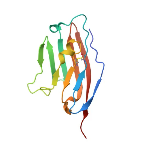
Monoclonal Antibodies
Anti-Mouse CD274/PD-L1 Monoclonal Antibody () for Flow Cytometry - Q9EP73
Item Number : CM0010283
Price varies based on specs and customizations
- Application
- FC
- Cross Reactivity
- Mouse
- Protein Weight
- 32kDa

Shipping:
Contact us to get shipping details Enjoy On-time Dispatch Guarantee.
Why Choose Us
Easy ordering process, quality products, and dedicated support for your business success.
Core Product Specifications and Parameters
| Parameter | Value |
|---|---|
| Product Name | Rabbit anti-Mouse CD274/PD-L1 mAb |
| Remarks/Alias | B7h1; Pdl1; Pdcd1l1; Pdcd1lg1; A530045L16Rik |
| Species | Mouse |
| GeneID (human) | 29126 |
| GeneID | 60533 |
| Immunogen | Recombinant fusion protein containing a sequence corresponding to amino acids 19-235 of mouse CD274/PD-L1 (NP_068693.1) |
| Source | Rabbit |
| Category | Monoclonal Antibodies |
| Application | FC |
| Cross Reactivity | Mouse |
| SWISS | Q9EP73 |
| Protein Weight | 32kDa |
| Shipping | Ice bag |
Biological Background: CD274/PD-L1 Function and Localization
- CD274, also known as PD-L1 or B7-H1, is a type I transmembrane protein belonging to the immunoglobulin superfamily.
- It acts as a ligand for the inhibitory receptor PDCD1/PD-1, playing a critical role in maintaining immune tolerance by limiting T-cell effector responses. Related references: PMID:11238124
- Through a yet undefined receptor, it may costimulate certain T-cell subsets that predominantly produce interleukin-10 (IL10). Related references: PMID:11015443 PMID:12719480
- Tumors exploit the PD-1/PD-L1 pathway to inhibit cytotoxic T lymphocyte (CTL) function and evade immune destruction. Blockade of this interaction can reverse T-cell exhaustion and enhance anti-tumor immunity. Related references: PMID:12218188 PMID:27281199
- PD-L1 localizes to the cell membrane and also cycles through early and recycling endosomes.
- It is highly expressed in heart, thymus, skeletal muscle, and lung, and is inducibly expressed on activated dendritic cells, B cells, macrophages, and various tumor cell lines.
- Post-translational modifications include N-linked glycosylation, disulfide bond formation, and ubiquitination, which modulate its stability and function.
Experimental Guidance and Technical Tips
- For flow cytometry, titrate the antibody to determine the optimal concentration for your specific cell type and experimental conditions.
- Use appropriate isotype controls and compensation controls when multiplexing with other fluorophores.
- Consider using Fc receptor blocking reagents to minimize non-specific binding in mouse samples.
- As the immunogen corresponds to the extracellular domain, the antibody is expected to recognize native surface PD-L1; live-cell staining is recommended for detecting surface expression.
- Although validated for flow cytometry, if applying to other techniques such as western blotting, note that glycosylation may cause an apparent molecular weight higher than the predicted 32 kDa; additional validation would be required.
CamelBio: Your One-Stop Sourcing Bridge
CamelBio is a premier sourcing partner for IVD raw materials, offering diagnostic manufacturers and research labs streamlined access to high-performance reagents. Our anti-PD-L1 monoclonal antibody addresses the growing demand for immune checkpoint targets in immuno-oncology assay development. With a portfolio that includes validated antibody pairs, optimized monoclonal and polyclonal antibodies, bulk ancillary reagents, and rare target sourcing, CamelBio supports the full diagnostic pipeline from concept to clinic.
REQUEST A QUOTE
Our professional team will reply to you within one business day. Please feel free to contact us!