Polyclonal Antibodies
Anti-LCK Rabbit Polyclonal Antibody for WB, IHC-P, ELISA - P06239
Item Number : CM0018747
Price varies based on specs and customizations
- Application
- WB, IHC-P, ELISA
- Cross Reactivity
- Human, Mouse, Rat
- Protein Weight
- 58kDa

Shipping:
Contact us to get shipping details Enjoy On-time Dispatch Guarantee.
Why Choose Us
Easy ordering process, quality products, and dedicated support for your business success.
Core Product Specifications and Parameters
| Parameter | Value |
|---|---|
| Product Name | LCK Rabbit pAb |
| Remarks/Alias | LSK; YT16; IMD22; p56lck; pp58lck; LCK |
| Species | Human |
| Gene ID (Human) | 3932 |
| Gene ID | 3932 |
| Immunogen | Recombinant fusion protein containing a sequence corresponding to amino acids 1-240 of human LCK (NP_005347.3). |
| Source | Rabbit |
| Category | Polyclonal Antibodies |
| Application | WB, IHC-P, ELISA |
| Cross Reactivity | Human, Mouse, Rat |
| SWISS | P06239 |
| Protein Weight | 58kDa |
| Shipping | Ice bag |
Biological Background: LCK Function and Localization
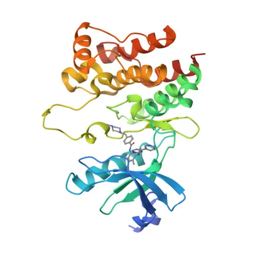
- LCK is a 56 kDa lymphocyte-specific tyrosine kinase (p56lck) and a member of the Src family. It contains SH2 and SH3 domains and is anchored to the plasma membrane via N-terminal myristoylation and palmitoylation.
- LCK is constitutively associated with the cytoplasmic tails of CD4 and CD8 co-receptors. Upon TCR engagement, LCK is recruited to the TCR-CD3 complex, where it phosphorylates tyrosine residues within the ITAMs of CD3 and TCRζ chains, initiating downstream signaling. Related references: PMID:2470098, PMID:40592325
- This kinase phosphorylates and activates ZAP70, leading to lymphocyte activation and lymphokine production. LCK also participates in IL-2 receptor signaling and phosphorylates other substrates including RUNX3, PTK2B, and RHOH. Related references: PMID:2470098
- LCK is expressed specifically in lymphoid cells and is present throughout thymocyte development, regulating both pre-TCR and mature αβ TCR maturation checkpoints.
- Subcellularly, LCK localizes to the cytoplasmic side of the cell membrane and in the cytosol, where it is poised to interact with surface receptors.
- Post-translational modifications include N-terminal lipid anchors (myristate and palmitate), multiple phosphorylation events (autophosphorylation at Tyr394 and inhibitory phosphorylation at Tyr505 by CSK), and ubiquitination. Alternate splicing produces isoforms.
- LCK is implicated in disease; chromosomal rearrangements involving LCK are associated with T-cell lymphomas, and germline mutations can cause severe combined immunodeficiency. Viral proteins such as HIV-1 Nef target LCK to modulate T-cell function.
Experimental Guidance and Technical Tips
- The immunogen (aa 1-240) covers the N-terminal unique and SH3 domains, which are less conserved among Src family members; this design may enhance specificity for LCK.
- For WB, a band at approximately 56-58 kDa is expected in lymphoid cell lysates (e.g., Jurkat, human PBMCs, mouse splenocytes). Optimize sample preparation and antibody dilution for your system.
- For IHC-P, antigen retrieval may be required; positive control tissues include human thymus or lymph node. Validate staining in relevant experimental samples.
- The antibody cross-reacts with mouse and rat LCK; if using other species, verify reactivity independently.
- As a polyclonal antibody, lot-to-lot consistency should be assessed; it is suitable for ELISA applications when paired with appropriate secondary antibodies.
CamelBio: Your One-Stop Sourcing Bridge
CamelBio is a specialized provider of IVD raw materials, offering diagnostic manufacturers and laboratories streamlined access to high-quality antibodies like this LCK rabbit pAb. Our portfolio includes validated antibody pairs, optimized monoclonal/polyclonal antibodies, and bulk ancillary reagents for immunology and oncology research. With flexible sourcing and technical support, CamelBio helps bridge the gap from target discovery to clinical assay deployment.
REQUEST A QUOTE
Our professional team will reply to you within one business day. Please feel free to contact us!