Co-existing structural variants like Hemoglobin E mask true HbA2 levels by co-migrating or co-eluting with it, causing a falsely elevated measurement.
This interference directly undermines the diagnosis of beta-thalassemia trait, where an accurate HbA2 value is the critical biomarker. The solution lies in methodologies that exploit different physicochemical properties—capillary electrophoresis (CE) and optimized cation-exchange HPLC—to achieve baseline resolution between HbA2 and common variant hemoglobins, a non-negotiable requirement for reliable IVD assay development and clinical reporting.
In diagnostic assay development, the presence of structural hemoglobin variants such as Hb E creates an analytical blind spot: they mimic HbA2 in traditional electrophoretic or poorly optimized chromatographic methods, leading to gross overestimation. The resolution comes from selecting separation technologies—specifically capillary electrophoresis and dedicated cation-exchange HPLC protocols—that separate molecules by both charge and hydrodynamic volume, turning a single, merged peak into two distinct, quantifiable zones.
How Structural Variants Disrupt HbA2 Quantification
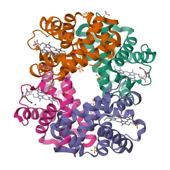
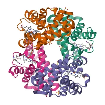
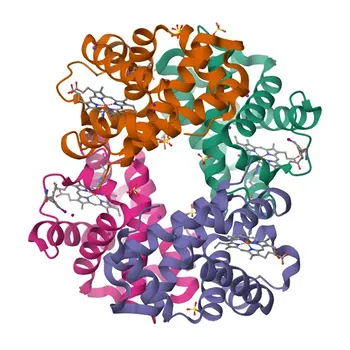
Diagnosis of beta-thalassemia trait hinges on elevated HbA2 (α2δ2). When a structural variant like Hb E (α2β2 with a β26 Glu→Lys substitution) is present, the analytical signal for HbA2 becomes compromised. The interference arises because the variant shares near-identical migration or retention characteristics with HbA2, making them look like a single entity.
The Co-Migration Trap in Electrophoresis
Standard alkaline cellulose acetate electrophoresis places Hb E exactly in the HbA2 band.
The two proteins have essentially the same overall charge-to-mass ratio under these conditions, so they move as one. The result is a densitometric trace that sums their signals, falsely inflating HbA2 and potentially triggering a false-positive thalassemia trait diagnosis in a simple heterozygous Hb E carrier.
The Co-Elution Problem in Routine HPLC
Not all HPLC methods are equal. Many commonly used ion-exchange HPLC systems (especially those using older or generic elution protocols) cannot differentiate HbA2 from Hb E.
Because both proteins elute within the same narrow retention-time window, they merge into a single peak. The integrator quantifies this combined area as "HbA2," producing an artifactually high value that obscures the true low/normal HbA2 level of an Hb E heterozygote.
The Biochemical Root of the Interference
HbA2 consists of α2δ2 chains, while Hb E is α2β2 with a point mutation in the β-chain. Their subtle differences in surface charge and hydrophobicity are insufficient to drive separation in many conventional systems.
This biochemical similarity forces assay developers to move beyond simple charge-based screening and to design or adopt methods that capitalize on finer molecular property differences.
Methodologies That Resolve the Interference
To design an IVD assay or choose a laboratory workflow that reports a true HbA2 value in the presence of Hb E, you must employ separation techniques that discriminate based on a combination of charge, molecular shape, and hydrodynamic radius. Two technologies stand out as definitive solutions.
Capillary Electrophoresis (CE): The Gold Standard for Resolution
Capillary zone electrophoresis (CZE) separates hemoglobin species inside a narrow fused-silica capillary under high voltage.
In CE, the electroosmotic flow and the electrophoretic mobility of each protein are influenced by both net charge and molecular size/shape. Hb E and HbA2 migrate at measurably different velocities, producing clearly separated peaks that enable accurate, individual quantification.
For an IVD developer, CE offers an orthogonal confirmation mechanism. Even if a primary HPLC method shows a borderline result, a CE run can cleanly split the HbA2 and Hb E zones, providing the true value without interference. The high resolution and automation compatibility make CE a favored platform for modern hemoglobinopathy screening.
Optimized Cation-Exchange HPLC: Separation Through Gradient Refinement
Cation-exchange HPLC separates proteins by their net positive charge binding to a negatively charged resin, eluting them with an increasing salt or pH gradient.
Unlike generic HPLC protocols, dedicated β-thalassemia programs on instruments like the Bio-Rad Variant II or Tosoh G8 use carefully tuned gradient slopes and column chemistries. These optimized methods exploit the slight charge differences between Hb E and HbA2 to yield distinct peaks—for example, HbA2 eluting at ~3.5 minutes and Hb E at ~4.0 minutes.
The key insight for assay development is that it’s not HPLC that fails, but unoptimized HPLC. When you build a cation-exchange method, you must validate the gradient and column selectivity using known Hb E-containing samples. If resolution is not achieved, you adjust buffer pH, gradient steepness, or column length until baseline separation is obtained. Only then is the method fit for diagnostic use.
Why Alkaline and Acid Electrophoresis Alone Are Insufficient
Traditional cellulose acetate electrophoresis (alkaline or acid) cannot be relied upon as the sole quantitation method when structural variants are suspected.
Alkaline electrophoresis merges Hb E with HbA2, and acid electrophoresis (citrate agar) may separate them but is primarily qualitative and unsuitable for precise percentage quantitation. Using these as standalone methods in a diagnostic kit leaves a dangerous blind spot.
Understanding the Trade-offs and Pitfalls
Every methodology comes with constraints that IVD developers and high-throughput labs must weigh before finalizing a workflow.
Cost and Throughput Considerations
Capillary electrophoresis instruments (e.g., Sebia Capillarys) require specific consumables and may have a lower sample throughput per hour compared to some high-speed HPLC systems.
Optimized HPLC analyzers can process hundreds of samples daily but require careful method maintenance and dedicated reagent lots. Budget, expected sample volume, and turnaround time demands will drive the final platform choice.
The Risk of Unrecognized Variants
Even with CE or optimized HPLC, rare variants with mobilities extremely close to HbA2 could still interfere. No technique is 100% immune.
Therefore, assay developers should build in a reflex algorithm: if an abnormal peak is detected in the HbA2 window, the system should flag the result and, if available, trigger a confirmatory CE run or molecular assay.
Reagent and Calibrator Consistency
Both CE and HPLC rely heavily on high-quality, purified hemoglobin controls (HbA2, HbA, HbF) and matched calibrators.
Lot-to-lot variability in column packing (HPLC) or capillary coatings (CE) can shift resolution. A robust IVD kit must include stabilized controls that challenge the separation of HbE from HbA2 in every run, ensuring the method remains interference-free over time.
Making the Right Choice for Your Diagnostic Goal
Your selection depends on whether you are developing a new IVD platform, validating a laboratory workflow, or interpreting patient results. Below are goal-oriented recommendations built on the principle that accurate HbA2 quantification demands verified separation of co-existing structural variants.
- If your primary focus is designing an IVD assay kit for hemoglobinopathy screening: Implement capillary electrophoresis as the core technology, or pair an optimized cation-exchange HPLC method with a reflexive CE system. Include a dedicated control material containing Hb E to validate resolution with each new reagent lot.
- If your primary focus is selecting a high-throughput clinical analyzer for a reference lab: Choose an automated HPLC platform with a verified β-thalassemia program that clearly resolves Hb E. Cross-validate at least 5% of all samples with abnormal HbA2 results using capillary electrophoresis to guard against subtle co-elution.
- If your primary focus is troubleshooting a current assay that gives falsely elevated HbA2 in Hb E carriers: Immediately review your HPLC gradient parameters or switch to a CE mode. Do not rely on post-hoc mathematical corrections; physically separate the species to get an artifact-free reading.
Your goal is not just a number, but a confident, interference-free measurement that directly protects patients from misdiagnosis. By embedding the resolving power of capillary electrophoresis or truly optimized cation-exchange HPLC into your workflow, you transform a common analytical pitfall into a solved problem.
Summary Table:
| Methodology | Separation Mechanism | Resolution Capability | Primary Application |
|---|---|---|---|
| Alkaline Electrophoresis | Net charge | Poor (Co-migrates with HbA2) | Basic qualitative screening only |
| Unoptimized HPLC | Ion-exchange charge | Poor (Co-elutes with HbA2) | High risk of false-positive HbA2 |
| Optimized Cation-Exchange HPLC | Fine surface charge gradient | High (Baseline separation) | High-throughput clinical screening |
| Capillary Electrophoresis (CE) | Net charge & hydrodynamic radius | Excellent (Gold standard) | IVD development & reflex validation |
Accelerate Your IVD Assay Development with CamelBio
Overcoming complex analytical interferences like co-existing structural hemoglobin variants requires robust methodologies and high-purity assay components. CamelBio provides diagnostic manufacturers, labs, and research institutes with one-stop access to IVD raw materials, technical services, and consulting—covering every stage from concept to clinic.
Whether you are refining HPLC gradients, setting up capillary electrophoresis protocols, or sourcing reliable hemoglobin standards, our technical team is ready to help you build interference-free diagnostic assays.
Contact CamelBio Today to enhance your assay precision and streamline your clinical diagnostic workflow.
Related Products
- Anti-HBA2 Rabbit Polyclonal Antibody for WB, IF-P, ELISA - P69905
- HABP2 Rabbit pAb for WB, IF/ICC, ELISA - Q14520
- Anti-Hemoglobin Subunit Alpha (HBA1) Monoclonal Antibody for WB, IF-P, IHC-P, ELISA - P69905
- Anti-Hemoglobin subunit alpha (HBA1) Rabbit Polyclonal Antibody for WB and ELISA - P69905
- Anti-HBB Polyclonal Antibody for WB, ELISA - P68871